Left Hip Muscles Anatomy - What Is A Hip Flexor Plano Orthopedic Sports Medicine Center : Rectus femoris muscle, one of.. The hip joint is an intricate structure including hip bones hip articular cartilage muscles ligaments and tendons and synovial fluid. These muscles are responsible for hip joint extension (backward movement). The hip is surrounded by thick muscles. The piriformis muscle is a key landmark in the gluteal region. It is also referred to as a ball and socket joint and is surrounded by muscles, ligaments, and tendons.
To put it plainly, sometimes hip pain comes from the hip, but a lot of times hip pain comes from the back. One at the left hip, and one at the right hip. The main action of the adductors is to pull the leg inward toward the other leg. Functionally, the hip joint enjoys a very high range of motion. These muscles include the gluteus maximus muscle (the largest muscle in the body) and the hamstrings group, which consists of the biceps femoris, semimembranosus, and semitendinosus muscles.
Anatomy Of Knee from marvel-b1-cdn.bc0a.com The deep gluteal muscles are a set of smaller muscles, located underneath the gluteus minimus. It works better during single movements. The inner thigh is formed by the adductor muscles. These muscles include the gluteus maximus muscle (the largest muscle in the body) and the hamstrings group, which consists of the biceps femoris, semimembranosus, and semitendinosus muscles. 1 hip anatomy, function and common problems. The strong muscles of the hip region also help to hold the hip joint together and prevent dislocation. The gluteals make up the muscles of the buttocks on the back of the hip. Thigh muscles also protect neurovascular structures as they go through the proximal hip joint to the knee and lower leg(3).
Knee assessment and hip mechanics online course:
The quadriceps group of four muscles. Ebraheim's educational animated video describes the muscle anatomy of the hip and buttocks region with simple images; Some of the other muscles in the hip are: These muscles are the adductor longus, adductor brevis, adductor magnus, gracilis, and the obturator externus. The main action of the adductors is to pull the leg inward toward the other leg. The medial thigh muscles are responsible for the adduction (movement of a body part toward the body's midline) of the leg. This mri hip joint axial cross sectional anatomy tool is absolutely free to use. These muscles include the gluteus maximus muscle (the largest muscle in the body) and the hamstrings group, which consists of the biceps femoris, semimembranosus, and semitendinosus muscles. 12 photos of the muscles of the lower back and hip diagram. This is because there are so many different muscles that give our hip joints a full range of motion. Your body has two iliopsoas muscles: The hip's essential muscles are the sartorius, rectus femoris, gluteus minimus and medius, iliopsoas, adductors, and hamstrings. The hip muscles are composed of multiple flexors, extensors, adductors, abductors, and rotators that work together.
The hip joint is an intricate structure including hip bones hip articular cartilage muscles ligaments and tendons and synovial fluid. These muscles are the adductor longus, adductor brevis, adductor magnus, gracilis, and the obturator externus. These ligaments reinforce and stabilize the hip joint(6). This mri hip joint axial cross sectional anatomy tool is absolutely free to use. One at the left hip, and one at the right hip.
Modified Osteotomy Of Posterolateral Overhanging Part Of The Trochanter Via Posterior Approach For Hip Arthroplasty An Anatomical Study Bmc Musculoskeletal Disorders Full Text from media.springernature.com The gluteals make up the muscles of the buttocks on the back of the hip. Weak adductor muscles may cause knee instability and adductor strain(2). Iliopsoas muscle, a hip flexor muscle that attaches to the upper thigh bone. 12 photos of the muscles of the lower back and hip diagram. Left hip muscles anatomy : One at the left hip, and one at the right hip. Adductor muscles on the inside of your thigh. 1 hip anatomy, function and common problems.
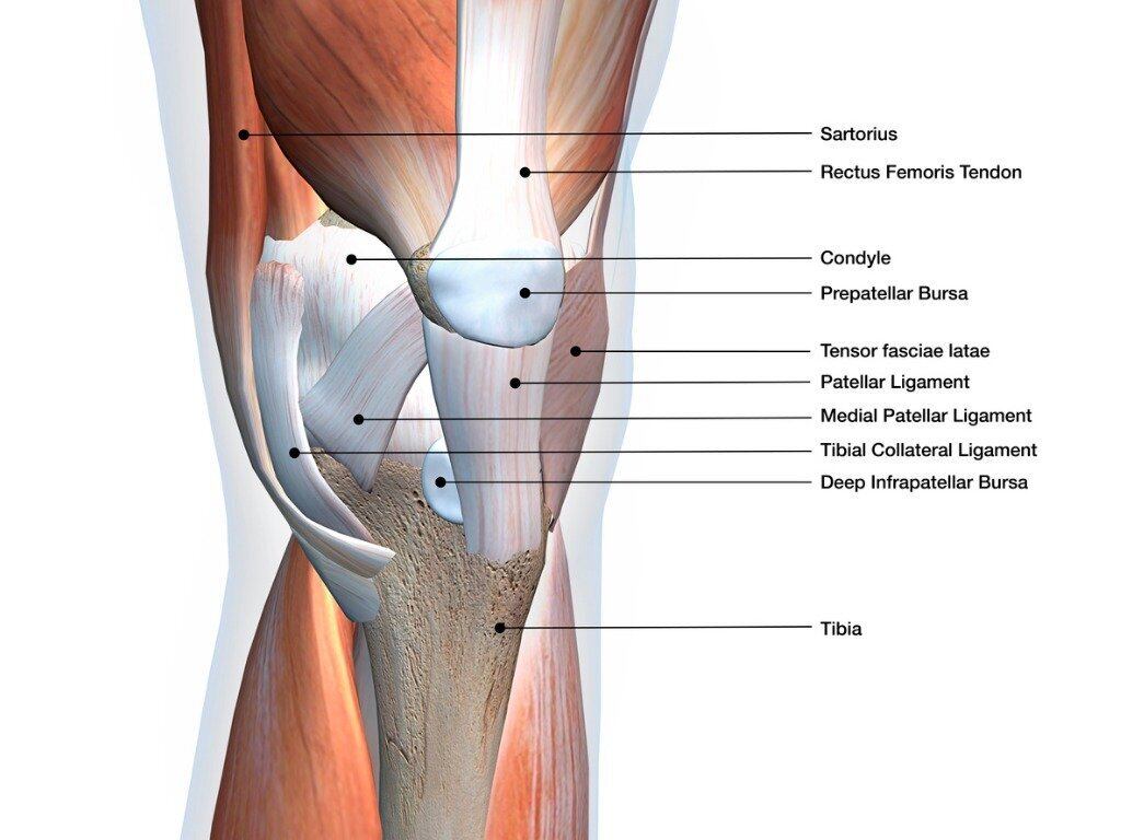
Anatomy of the hip muscles.
The hip muscles are composed of multiple flexors, extensors, adductors, abductors, and rotators that work together. These muscles include the gluteus maximus muscle (the largest muscle in the body) and the hamstrings group, which consists of the biceps femoris, semimembranosus, and semitendinosus muscles. Muscles of the gluteal region: The inner thigh is formed by the adductor muscles. Rectus femoris muscle, one of. They also stabilise the hip joint by 'pulling' the femoral head into the acetabulum of the pelvis. Pick which works for you and then. These muscles are responsible for hip joint extension (backward movement). The quadriceps group of four muscles. Blood vessels and nerves of the hip They begin under the gluteus maximus behind the hipbone and attach to the tibia at the knee. This is because there are so many different muscles that give our hip joints a full range of motion. The hamstrings are three muscles at the back of the thigh that affect hip and knee movement.
Use the mouse scroll wheel to move the images up and down alternatively use the tiny arrows (>>) on both side of the image to move the images.>>) on both side of the image to move the images. The quadriceps group of four muscles. The femur may also rotate around its axis about 90 degrees at the hip. Muscles of the gluteal region: 1 hip anatomy, function and common problems.
Hip Pain 13 Year Old Female from pnpfitness.com The piriformis muscle is a key landmark in the gluteal region. In utero fetal hips lie typically in flexion, abduction and external rotation, with the left hip usually muscular anatomy. Knee shoulder shoulder arthrogram ankle elbow wrist hip contact. The hamstrings are three muscles at the back of the thigh that affect hip and knee. The hip is surrounded by thick muscles. Rectus femoris muscle, one of. These muscles are the adductor longus, adductor brevis, adductor magnus, gracilis, and the obturator externus. The hip muscles are composed of multiple flexors, extensors, adductors, abductors, and rotators that work together.
This mri hip joint axial cross sectional anatomy tool is absolutely free to use.
This mri hip joint axial cross sectional anatomy tool is absolutely free to use. Left hip muscles anatomy : Pick which works for you and then. The medial thigh muscles are responsible for the adduction (movement of a body part toward the body's midline) of the leg. In utero fetal hips lie typically in flexion, abduction and external rotation, with the left hip usually muscular anatomy. Injury to the iliopsoas may cause hip pain and limited mobility. Hip muscle anatomy is a complex topic. Posterior view of gluteus maximus and gluteus medius in human anatomy, the muscles of the hip joint are those muscles that cause movement in the hip. The general action of these muscles is to laterally rotate the lower limb. What is collectively referred to as the hip flexors is actually a group of muscles that includes the iliopsoas, the thigh muscles (rectus femoris, sartorius and tensor. Rectus femoris muscle, one of. Adductor muscles on the inside of your thigh. Pelvis and acetabulum, with muscle attachment sites.


0 Komentar